MEDICAL PHYSIOLOGY

DISCUSSION FORUM IN TELEGRAM

MEDICAL PHYSIOLOGY/b>

Click here to access Unit one Content..


In order to function, cells require food.  On the other hand cell metabolism produces waste products that must be eliminated into the extracellular environment. 

Because the plasma membrane separates the intracellular fluid (ICF) from the extracellular fluid (ECF),

 All substances that either enter or leave the cell must pass through the plasma membrane

•      The membrane also keeps valuable proteins and other substances from leaving the cell and prevents the admission of undesirable substances.  For this reason; the cell membrane is classified as a semipermeable membrane.  (i.e it is permeable or passable to some molecules but impassable to others).

•      In general the mechanisms of cellular exchange can be divided into two categories, Active and Passive

•      Active require the cell to expend energy (ATP) to effect solute movement across the plasma membrane, whereas passive mechanisms do not require energy expenditure by the cell.

Passive transport

Osmosis

The diffusion of water through a semi-permeable membrane, with the water molecules going from the less concentrated solution to the more concentrated solution.

The dissolved molecules in the more concentrated solution ‘pull’ the water molecules from the less concentrated solution across the semipermeable membrane.

Hypertonic fluids 

Contain higher concentration of osmotically active particles than blood plasma.


Hypotonic solutions

Contain a low concentration of osmotically active particles than plasma.

Osmosis is important in maintaining plasma volume, interstitial and intracellular fluid volumes and the other fluid compartments.

When the body retains salt, large amounts of water are also retained.

Isotonic fluids

Contain – Osmotically active particles in the same concentration as found in the plasma of blood.  e.g.  0.9% Sodium Chloride in water.

Diffusion

•      The constant movement of molecules from a region of relatively higher concentration to one of lower concentration.

•       If the substance is equally distributed between two regions, no concentration gradient exists and diffusion equilibrium is present.

•       Movement is controlled by interruptions in the membrane called channels or pores.  Pores are fluid-filled channels formed by proteins within the membrane. 

An example of Diffusion

The movement of oxygen molecules into cells and carbon dioxide molecules out of cells oxygen molecules are being continually consumed by metabolic processes occurring within the cell, so that the concentration gradient favors diffusion of this gas into the cell.

•       Carbon dioxide molecules are being continually produced during cellular metabolism, so that the concentration gradient favors diffusion of this gas out of the cell.

Facilitated diffusion

•      This is aided by carrier proteins -e.g. integral proteins/ globular proteins act as carriers that transport materials in the opposite direction against their natural direction.

•      These carrier proteins are distinguished by two characteristics:-

•      Selectivity

•      Gates 

Selectivity

•      Due to characteristic of channel such as diameter, shape and nature of electrical charges on the inner surface.

Example sodium channels are 0.3 by o.5 nanometer and the fact that the inner surface is negatively charged.

•      Potassium channels are 0.3 by 0.3

Gates

•      Channels are opened and closed by gates.

•      The opening and closing of gates are controlled by two principal ways;

Voltage gating :

 This is where the gate responds to the electric potential across the cell membrane.

Ligand gating :

Caused by the binding of another molecule with the protein, this causes conformational change in the protein leading to opening or closing of the gates is referred to as ligand gating.

E.g. acetylcholine an excitatory neurotransmitter. it allows movement of positively charged sodium ions and transmission of impulse.(enables impulse to leap junction) 

Factors that may affect diffusion

Permeability of membrane due to;

•      Thickness of membrane

•      Lipid solubility

•      Number of protein channels

•      Temperature- increased temperature. Causes increased rate of diffusion.

•      Molecular weight of a substance

Concentration difference of diffusing substance.

Effect of electrical potential on diffusion of ions i.e. between the two sides of the membrane

The pressure difference across the membrane movement of substance occur from high to low pressure

Filtration

•      Filtration, the passage of water containing dissolved materials through a membrane as a result of a mechanical force on one side.

•      E.g. of filtration in the human body is the formation of urine in the microscopic functional units of the kidney.

Active Transport

Often molecules move into or out of living cell in an opposite direction from the way in 

•      which they would normally flow by diffusion

•       I.e. move from an area where they are in relatively lower concentration.

•      This movement because it is against the natural flow, requires energy in the form of ATP. It also requires proteins in the cell membrane that act as carriers for the molecules.

•      This process allows the cell to take in what it needs from the surrounding fluids and to release materials from the cell.

•      Cell membrane is described as selectively permeable based on the needs of the cell.

Endocytosis and Exocytosis

•      These are methods of bringing particles into the cell and releasing secretions to the exterior of the cell.

Endocytosis refers to the bringing in of protein and other substances through invagination of the outer cell membrane.  The process occurs in the following ways:

Pinocytosis (Solution) / “Cell drinking”

•      Involves movement of complex proteins and some strong electrolyte solutions into the cell. 

•      The protein is seen to adhere to the outer cell membrane, which stimulates invagination of the membrane.

•      The material is enclosed in a vesicle and floats into the cytoplasm.

•      Lysosomes attach to the vesicle, surface, release hydrolytic enzymes into the vesicle, and the enzymes break down the complex material for use within the cells.  Substances ingested are in solution hence not visible under the microscope.

Phagocytosis ‘cell eating’

•      The process by which bacteria, dead tissue or other bits of material visible under the microscope are engulfed by cells such as the polymorhonuclear leucocytes of the blood.

The material makes contact with the cell membrane, which then invaginates.  The  invagination is pinched off, leaving the engulfed material in the membrane – enclosed vacuole and the cell membrane intact.

Exocytosis (“Emeocytosis – (“Cell vomiting”)

•      This has been called reverse pinocytosis and is an active release of soluble products to the ECF.

•      Proteins that are secreted by cells move from the endoplasmic reticulum to the Golgi complex.  Where they are packaged into secretory granules..

•      To be secreted the granules adhere to the inner cell membrane.  The area of fusion breaks leaving the contents of the granule outside the cell and the cell membrane intact. EXAMPLE-secretion of waste products from cell, or release of neurotransmitters.

•      NB Both endocytosis and exocytosis require energy and are affected by cellular ability to synthesize ATP.

The Sodium-Potassium pump

Referred to as Sodium-Potassium –activated adenosine triphosphatase (Naa-K+ Atpase). 

It’s an active transport mechanism which pumps sodium outward through the cell membrane and at the same time pumps potassium from the outside to the inside.

•      The Sodium-Potassium pump responsible for the coupled active transport of Na+ out of cells and K+ into cells is a unique protein in the cell membrane.

•      This protein is also an adenosine triphosphatase i.e. an enzyme that catalyze the hydrolysis of ATP to Adenosine diphosphate (ADP) its activated by Na+ and K+.  

•      The pump extrudes 3Na+ from cells for each two K+ it takes into the cell. Ration3/2.The carrier protein (molecular weight 100,000)

The carrier protein (molecular weight 100,000), Has 3 receptor sites for binding to Na+ on  a part that protrudes to the interior cell. Has 2 receptor sites for K+ binding on the outside.

•      The inside portion of this protein (near the Na+ binding site) has an Atpase activity.

•      The amount of Na+ extruded by cell is partly regulated in a feedback by the amount of Na+ in the cell. When 3Na+ bind on the inside of the carrier protein and 2K+ on the outside the Atpase becomes activated

•      This cleaves one molecule of ATP to split to ADP –liberating energy which aid in in extruding Na+ to the outside and potassium to the inside.

Functions of the sodium-potassium pump

•      It’s present in all cells of the body.

•      Responsible for establishing a negative electrical potential inside the cells.

•      It is the basis of Nerve function to transmit nerve signals across the nervous system.

•      The Sodium-potassium pump contributes to the membrane potential.

•      controls cell volume, regulates water volume (osmosis of water to interior cell may lead to cell burst this is prevented by the movement 3 sodium that moves together with water to  the exterior of the cell.

Resting  membrane potential in transport

  Resting membrane potential (Steady potential.)

There is a potential difference across the membranes of most of cells, with the inside of the cells Negative to the exterior there are channels via which ions pass

•      The channels for Na+ K+ and Cl are all different.

•       The Sodium-potassium pump contributes to the membrane potential.

•       It maintains the concentration gradient on which the membrane potential depends.

•      If the resting membrane potential is decreased by the passage of a current through the membrane, the electrical gradient that keeps K+ inside the cell is decreased, and there is an increase in K+ diffusion out of the cell.


Click here to access Unit Two Content..


Topic 4: FLUIDS ,ELECTROLYTE BALANCE AND REGULATION OF BODY TEMPERATURE

Topic objective

1. Explain fluid and electrolyte balance in the body

2. Explain the regulation of body temperature

FLUIDS AND ELECTROLYTE BALANCE.

Review of homeostasis (Student discuss)

This is the steady state within an organism – it means staying the same.(Homeo- the same) and (Stasis – Staying)

•      Homeostasis involves maintenance of constant conditions in the internal environment.

 Homeostasis is process by which a stable internal environment is maintained despite changes in the external environment.

•   Homeostasis is maintained primarily by negative feedback systems – which means – The information is the system is used to reverse changes in the system to bring it back to normal. N/B positive feedback-is more of vicious circle. Useful in blood clotting or delivery.

Homeostatic control mechanism is  maintained Feedback Mechanism

•      Negative feedback

•      Positive feedback

Negative feedback: Response triggered by changed conditions serves to reverse the change

•      E.g., Body temperature increases à Skin blood vessels dilate à Body temperature decreases

Positive feedback occurs when the outcome of a process increases or intensifies that process.

§  During childbirth, hormones cause muscles of uterus to contract

§  Uterine contractions cause even more hormones to be released, which intensify the contractions

 In animals, regulation is usually by negative feedback because positive feedback often results in amplification – away from 

Regulation of water

Water is essential for fluids balance in the body

•       Water balance refers to an equilibrium maintained between intake and output. Water accounts for approximately 60% of body weight in a 70kg average male.

•      The amount of water necessary to maintain life per a day is 1500ml.

Factors that contribute to regulation of water.

Thirst

•      Thirst is the principal regulator of water intake. Osmoregulators in the thirst center in the hypothalamus are sensitive to changes in the osmolality of the extracellular fluids. Thirst may also be induced by the following;

•      Reduced circulatory volume due to hemorrhage.

•      Maybe induced by dryness of the mouth in true hyperosmolar states or may occur to relieve the unpleasant dry sensations that result from reduced salivation.

Renal regulation

•      Kidneys regulate the volume and electrolyte concentration of body fluids through:

•       Glomerular filtration

•      Tubular reabsorption

•      Tubular secretion and excretion

Antidiuretic hormone

•      The major stimuli for ADH secretion are increased osmolality, and decreased volume of extracellular fluid.

•      This hormone increases reabsorption of water at the collecting ducts thereby conserving water to correct osmolality and restore the volume of extracellular fluid

Aldosterone(Secreted by the adrenal glands)

•      Acts on the renal tubules to increase the sodium uptake.

•      Increased sodium retention causes increase in water retention.

Prostaglandins

naturally occurring fatty acids present in many tissues of the body

•      In kidneys renal prostaglandins cause vasodilatation promoting sodium excretion by inhibiting the responses of renal distal tubules to ADH

Gluco-corticoids

Exert weak mineral corticoid (aldosterone) activity thus promoting the resorption of sodium and water.

Electrolytes 

Electrolytes are compounds that form ions whenever they are in solution. Electrolytes are responsible for acidity and alkalinity of solutions.

Electrolytes and their functions

Sodium-

It is the chief responsible for maintaining osmotic balance and body fluid volume.

It is the main positive ion in extracellular fluids sodium is required for nerve impulse conduction and is important in maintaining acid base balance

Potassium

Important in the transmission of nerve impulses and is a major positive ion in intracellular fluids.

Involved in cellular enzyme activities, helps regulate the chemical reactions by which carbohydrate is converted to energy and amino acids to proteins.

Calcium-

Required for bone formation, muscle contraction, Nerve impulse transmission and blood clotting.

Phosphate-

Essential in metabolism of carbohydrates, bone formation and Acid-base balance. Phosphates are found in the cell membrane and the DNA

Chloride-

Is essential for formation of the hydrochloric acid of the gastric juice

Magnesium-

 Found mostly within the cells and in the bones. Activates a number of intracellular enzyme systems, is required for protein and nucleic acid synthesis, also particularly essential in promoting neuromuscular integrity.

Electrolytes are important in acid base balance

•      The ph. of the body must be held within a normal range at appprox.7.4.0

systems involved in maintaining acid base balance-;

Buffers –are substances that help maintain a constant ph by accepting or releasing hydrogen ions (H+)

•      The main buffer systems in the body are bicarbonate buffer phosphate buffers and proteins, such as hemoglobin and plasma proteins

The kidneys –serve to regulate PH by reabsorbing or eliminating hydrogen ions as needed much of the hydrogen that is eliminated by the kidneys is transported into the nephron by tubular secretion

Role of respiration-in controlling ph. release of carbon dioxide from lungs acts to increase the PH of the blood by reducing the amount of carbonic acid formed. A reduction in the release of carbon dioxide acts to decrease the ph. of the blood .Drop in ph. leads to acidosis while increase in ph. results in alkalosis.

REGULATION OF BODY TEMPERATURE

Heat production-heat produced when oxygen combines with products in the cells. The amount of heat produced by a given organ varies with the kind of tissue and its activity.

•      In rest muscles may produce 25% of total body heat but when muscles contract it may be a hundred times more owing to increase in metabolic rate.

•      In rest abdominal organs particularly liver produce about 50% total heat, and brain 15% of body heat.

•      The largest amount of heat ,therefore is produced in the muscles and glands

•      The circulating blood distributes the heat evenly.

Factors that affect heat production include:-

•      Activity level

•      Hormone production e.g. thyroxin and adrenaline

•      Food intake-readily available for cellular metabolism. The glandular structures and the muscles of the digestive system generate additional heat as they set to work.

•      Age.

Heat loss-Heat is lost through-

•      Skin 80%

•      Respiratory system and with urine and feces (15 to 20%)

•      Dissipation to the outside through- conduction, radiation, convection and evaporation.

Excess heat loss is prevented by-;

•      Artificial- keeping warm by clothes or warm air.

•      Natural-layers of fat under the skin prevent deeper tissues from losing much heat. Volume of tissue compared with the amount of skin surface .e g Child loses heat more rapidly than an adult

How is temperature regulated?

Heat regulation center is hypothalamus - Has receptors for heat loss and others for heat production. Which respond to –;

•      Heat brought to the brain by the blood

•      Nerve impulses from temperature receptors in the skin

If these two factors indicate that too much heat is being lost impulses are sent to the hypothalamus to the ANS which in turn causes-

•      Constriction of skin vessels in order to reduce heat loss

•      Impulses are sent to muscles to cause shivering, this is caused by rhythmic contraction of many body muscles resulting into increased heat production.

•      Epinephrine  (adrenal gland) production increases heat production by increasing cellular metabolism

•      Smooth muscle around the hair roots contract forming goose flesh.(goose pimples)


Hypothalamus Regulation of Temperature

•      Mechanisms are activated in two ways:

–     Thermal receptors in skin provide input to central command

–     Direct stimulation of hypothalamus through changes in blood temperature perfusing area

If these two factors indicate that there is too much heat than necessary -impulses are sent to the hypothalamus to the ANS which in turn causes-

•      Stimulate the sweat glands to increase their activity.

•      Causes dilation of blood vessels in the skin that there is increased blood flow and loss of heat

•      Causes relaxation of muscles and thus minimize the production of heat

Hormones  in temperature regulation

•      Antidiuretic hormone (ADH) is released to increase water re-absorption from kidneys.

•      Aldosterone is released to increase the re-absorption of sodium.

•      thyroxine –increase metabolism

Example

Negative feedback control of thyroxin in heat regulation


Circulatory Adjustments.

•      Cardiovascular drift – fluid loss reduces plasma volume (about 10% of fluid lost comes from plasma. About 50% comes from intracellular water).

•      Visceral vascular constriction and skin & muscle vascular dilation.

•      Maintaining blood pressure. Circulatory regulation and maintenance of muscle blood flow take precedence over temperature regulation often at the expense of spiraling core temperature during exercise in heat.

NOTE

•      Very young and very old persons are limited in their ability to regulate body temperature.

•       Normal body temperature is 36.2 to 37.60c but varies with time of the day.

Abnormal temperature

Fever –temperature high than normal can be caused by-;

•      Infections

•      Malignancies

•      Brain injuries

•      Toxic reactions reaction to vaccines

•      Diseases involving CNS

 Excessive heat exposure leads to-heat cramps, heat exhaustion, and heat stroke

 Excessive cold exposure leads to-hypothermia, frostbite, reduction of blood supply and tissue damage


Click here to access Unit Two Content..


Topic 4: Summary

*      Core temperature is a dynamic equilibrium as a result of balance between heat gain and heat loss.

*      Mean body temperature represents an average of skin and internal temperatures.

In Cold

*      Vascular adjustments: constrict peripheral blood vessels.

*      Muscular activity: exercise energy metabolism and shivering.

*      Hormonal output: epinephrine and norepinephrine increase basal heat production; prolonged cold – thyroxin.

When  it is hot

*      Heat transferred as water is vaporized from respiratory passages and skin surfaces.

*      For each liter of water vaporized, 580 kcal transferred to the environment.

*      When sweat comes in contact with the skin, a cooling effect occurs as sweat evaporates.

*      The cooled skin serves to cool the blood.


Click here to access Unit Three Content..


Topic 1: Functions of blood:

Functions of blood:

  1.  Blood fulfils a communication role between cells.  Thus, it is vital that each cell is in close communication with a blood vessel.
  2. Blood carries O2 from the lungs to the tissues and CO2 from the tissues to the lungs for disposal.
  3. Transport of other waste products for disposal.  For example to the kidneys for excretion, or to the liver for metabolism.
  4. Endocrine gland secretions (hormones) are carried to target tissues.
  5. Blood provides an immunological function.
  6. Thermoregulation.  Heat generated by muscles is carried to the skin.

Summary of blood functions:

 Distribution of :-

  1. O2 and nutrients to body cells
  2. Metabolic wastes to the lungs and kidneys for elimination
  3. Hormones from endocrine organs to target organs

  Regulation role:-

  1.      Body temperature by absorbing and distributing heat
  2.       Normal pH using buffers
  3.        Adequate fluid volume in the circulatory system

 Protection against:-

  1.     Blood loss
  2.     Plasma proteins and platelets initiate clot formation
  3.     Infection
  4.     Antibodies
  5.     Complement proteins
  6.     WBCs defend against foreign invaders

Physical characteristics of blood    

  1.  Sticky, opaque fluid
  2. Color scarlet to dark red
  3. pH 7.35–7.45
  4. 38°C
  5. ~8% of body weight
  6. Average volume: 5–6 L for males, and 4–5 L for females

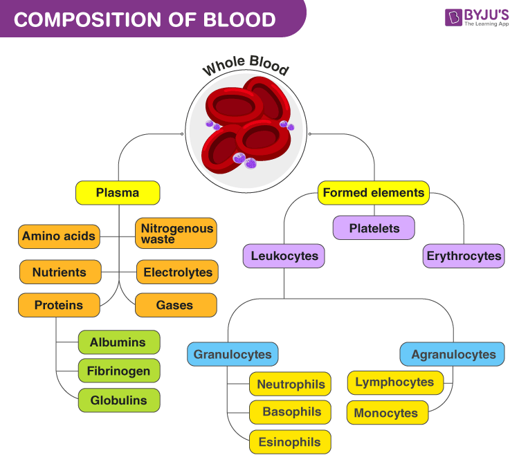
Blood: a fluid connective tissue composed of

¨      Plasma  

¨      Formed elements:

¨      Erythrocytes (red blood cells, or RBCs)

¨      Leukocytes (white blood cells, or WBCs)

¨      Platelets

 Hematocrit:

¨      Percent of blood volume that is RBCs

¨      47% ± 5% for males

¨      42% ± 5% for females

Blood  Plasma: Plasma is a solution containing :

  1. Water 90%,
  2.  Proteins: - albumin, globulins and fibrinogens most produced by the liver.
  3.   ions and small molecules
  4. Nitrogenous by-products of metabolism—lactic acid, urea, creatinine
  5. Nutrients—glucose, carbohydrates, amino acids
  6. Electrolytes—Na+, K+, Ca2+, Cl, HCO3
  7. Respiratory gases—O2 and CO2
  8. Hormones

  • Plasma protein:


Albumin:

 Predominant protein in blood. (60%).
Produced by the liver at a rate of synthesis = 14-15 g daily.
Its concentration is dependent upon nutritional status, particularly the amino acids.


  Globulins :( 36%).

  • Classified into 3 main types:a ,b and g globulins
  • many of the proteins in the plasma e.g. transferrin, complement, enzymes and carriers are a and b globulins
  • a and b globulins are produced by the liver
  • g Globulins are immunoglobulins (antibodies).
  • g globulins are produced by the plasma cells

        Fibrinogen:

         forms the blood clot
         Produced by the liver


Functions of plasma proteins

  1. Protein reserve in nutritional depletion
  2. Transporter: e.g. of fatty acids ,bilirubin
  3. Binding and solubilization of drugs
  4. Regulator of colloidal pressure

NB: Decreases in albumin concentration cause edema.
        Causes of decreased albumin/abnormal excretion/degradation of albumin: - Malnutrition, Advanced chronic liver disease, nephrotic syndrome, burns.


Click here to access Unit Three Content..


Topic 2: Haemopoiesis. (Production of blood cells).

Haemopoiesis. (Production of blood cells).

    1.  Production of all blood cells takes place only in the bone marrow after birth.
    2. During the intrauterine life the liver, spleen and yolk sac produce blood cells
    3. The marrow in all the bones contribute in the haemopoesis up to age of 20 years
    4. After 20 years haemopoesis is confined to the marrow of the flat bones and ends of long bones.
    5. The marrow that continue production is called the red marrow
    6. The marrow in the shaft of long bones is converted into fatty tissue and is called the yellow marrow.
    7. All blood cells are derived from a single type of cell called pluripotent stem cells mainly the :-
      1. Myeloid progenitor which forms the erythrocytes, neutrophils and platelets.
      2. Lymphoid progenitor which forms B- lymphoid and T-lymphocytes.

Red blood cells.( Erythrocytes)

  •  Biconcave discs, no nucleate, essentially no organelles.
  • Filled with hemoglobin (Hb) for gas transport
  • Contain the plasma membrane protein spectrin and other proteins

¨      Provide flexibility to change shape as necessary

  • Are the major factor contributing to blood viscosity

Development of RBC. (Erythropoiesis).

  • The first recognized red cell precursor is called pronormpblast:

·         Pronormoblast undergoes change in size, colour and contents to reach the mature RBC and passing through the following stages:

·         Pronormoblast

·         Early normoblast

·         Intermediate normoblast

·         Late normoblast

·         Reticulocyte

·         Mature RBC

  • Some reticulocytes enter blood before full maturation
  • 30% of the Hb is formed in the reticulocytes
  • It takes 2 days to develop into mature RBC
  • Reticulocyte count in blood is 1%
  • Reticulocytes count is an indicator of bone marrow activity
  • An increase in the reticulocyte count (reticulocytosis) indicates increase bone marrow activity e.g. in hemolytic anemia
  • The mature RBC has no nucleus or other organelles
  • It is a bag of haemoglobin
  • A biconcave disc containing haemoglobin
  • Cells with volume < 80 :microcyte
  • Cells with volume >95 :macrocyte

Importance of the biconcave shape:

  • Increases surface area
  • Increases flexibility
  • Decreases fragility
  • Cells with an abnormal shape breaks down easy
  • Patients with hereditary spherocytosis they have anaemia due to excessive breakdown of the red cell.
  • Fragility of the cells can be tested by osmotic fragility test

Control of Red Cell Production:

  • Normal RBC count is 5 million /mm3
  • Count is higher in males than females. Testosterone enhances EPO production, resulting in higher RBC counts in males.
  • Red cell function is to transport O2 to tissues
  • O2 need by the tissues is the main regulator of RBC production

Regulation of RBC production:

Kidney?

Erythropoeitin?  +

Bone marrow?

Red blood cell?

Oxygen?


An increase in RBC count is called polycythaemia.

    • Physiological polycythaemia as occurs at high altitude in response to decrease in O2 pressure
    • Pathological polycythaemia: as for example a tumor of the kidney producing large amounts of Erythropoeitin
    • Effects of polycythaemia:- Increases the blood viscosity

Hemoglobin

    • Found inside the RBC
    • Main function is O2 transport
    • Contributes to the buffering capacity of the blood

Structure:

Consists of 2 parts: Globin and Haem. Hemoglobin consists of globin (two alpha and two beta polypeptide chains) and four heme groups.


Haemoglobin – Part 1 – Hemoglobin (Hb) – Labpedia.net


 Globin

    • A protein that consists of 4 polypeptide chains.
    • According to the type of chain HB is classified into:
  1.  -Hemoglobin A: forms 98.5% of the hemoglobin. It of consists of 2a and 2b

  2. -Hb A2: Consists of 2a and 2d . Forms about 1.5% of Hb in adults.

  3. -Hemoglobin F: the haemoglobin of the foetus. Consists of 2a and 2g . Haemoglobin F has higher affinity to O2 than Hb A. This facilitates the transfer of O2 from the mother to her baby.

          The Haem

      • It is an iron containing pigment
      • The pigment is a porphyrin ring
      • Hb contains 4 haems
      • The iron is in the reduced form(ferrous)
      • O2 is transported by Hb bound to iron
      • The binding is weak and does not lead to oxidation of the iron
    •        

      • Haemoglobin+O2      ?        oxyHb
      • Haemoglobin+CO2      ?           carbaminoHb
      • Haemoglobin +CO       ?        carboxyHb
      • CarboxyHb cannot transport O2
      • Affinity of CO to bind to Hb is x200 than O2
      • CO is a toxic gas
      • Hb + Glucose ?Glycosylated Hb.(Hb1Ac) - The level of HbA1c is a good indicator of control of diabetes mellitus

    •        Reactions of haemoglobin:

        1. Haemoglobin+O2      ?        oxyHb
        2. Haemoglobin+CO2      ?           carbaminoHb
        3. Haemoglobin +CO       ?        carboxyHb
        4. CarboxyHb cannot transport O2
        5. Affinity of CO to bind to Hb is x200 than O2
        6. CO is a toxic gas
        7. Hb + Glucose ?Glycosylated Hb.(Hb1Ac) - The level of HbA1c is a good indicator of control of diabetes mellitus

      • Haemoglobinopathies:

        • Abnormal Hb due to abnormality in the globin part:

        Haemoglobin S:

        1. In the b  chain at position No 6 glutamic acid is replaced by valine
        2. The change may affect one or both b chains
        3. The condition is hereditary
        4. It is common in Africa
        5. Hb S is less soluble especially when exposed to low O2 tension and precipitates in the cell
        6. Precipitation of the Hb inside the cell>>>sickle shape
        7. The sickle cells  break down easier
        8. When only one b chain is abnormal>>sickle cell trait
        9. When the two b chains are abnormal>>>sickle cell anemia.

               Thalassemia:

        The No of chains are less
         One or both a  chains may be missing>>> a thalassaemia
         One or both b  chains may be missing>>> b  thalassaemia
         The affected cells have abnormal shape and breaks down easier>>>anaemia
        Thalassaemia is common in the Mediterranean area.

             Dietary Factors Needed for Normal erythropoiesis

                      Iron

                    vitamin B12

                    folic acid

                    More important to women due to the loss of blood during menstruation, children and young girls.


Click here to access Unit Three Content..


Topic 2: Haemopoiesis. (Production of blood cells).

Haemopoiesis. (Production of blood cells).

    1.  Production of all blood cells takes place only in the bone marrow after birth.
    2. During the intrauterine life the liver, spleen and yolk sac produce blood cells
    3. The marrow in all the bones contribute in the haemopoesis up to age of 20 years
    4. After 20 years haemopoesis is confined to the marrow of the flat bones and ends of long bones.
    5. The marrow that continue production is called the red marrow
    6. The marrow in the shaft of long bones is converted into fatty tissue and is called the yellow marrow.
    7. All blood cells are derived from a single type of cell called pluripotent stem cells mainly the :-
      1. Myeloid progenitor which forms the erythrocytes, neutrophils and platelets.
      2. Lymphoid progenitor which forms B- lymphoid and T-lymphocytes.

Red blood cells.( Erythrocytes)

  •  Biconcave discs, no nucleate, essentially no organelles.
  • Filled with hemoglobin (Hb) for gas transport
  • Contain the plasma membrane protein spectrin and other proteins

¨      Provide flexibility to change shape as necessary

  • Are the major factor contributing to blood viscosity

Development of RBC. (Erythropoiesis).

  • The first recognized red cell precursor is called pronormpblast:

·         Pronormoblast undergoes change in size, colour and contents to reach the mature RBC and passing through the following stages:

·         Pronormoblast

·         Early normoblast

·         Intermediate normoblast

·         Late normoblast

·         Reticulocyte

·         Mature RBC

  • Some reticulocytes enter blood before full maturation
  • 30% of the Hb is formed in the reticulocytes
  • It takes 2 days to develop into mature RBC
  • Reticulocyte count in blood is 1%
  • Reticulocytes count is an indicator of bone marrow activity
  • An increase in the reticulocyte count (reticulocytosis) indicates increase bone marrow activity e.g. in hemolytic anemia
  • The mature RBC has no nucleus or other organelles
  • It is a bag of haemoglobin
  • A biconcave disc containing haemoglobin
  • Cells with volume < 80 :microcyte
  • Cells with volume >95 :macrocyte

Importance of the biconcave shape:

  • Increases surface area
  • Increases flexibility
  • Decreases fragility
  • Cells with an abnormal shape breaks down easy
  • Patients with hereditary spherocytosis they have anaemia due to excessive breakdown of the red cell.
  • Fragility of the cells can be tested by osmotic fragility test

Control of Red Cell Production:

  • Normal RBC count is 5 million /mm3
  • Count is higher in males than females. Testosterone enhances EPO production, resulting in higher RBC counts in males.
  • Red cell function is to transport O2 to tissues
  • O2 need by the tissues is the main regulator of RBC production

Regulation of RBC production:

Kidney?

Erythropoeitin?  +

Bone marrow?

Red blood cell?

Oxygen?


An increase in RBC count is called polycythaemia.

    • Physiological polycythaemia as occurs at high altitude in response to decrease in O2 pressure
    • Pathological polycythaemia: as for example a tumor of the kidney producing large amounts of Erythropoeitin
    • Effects of polycythaemia:- Increases the blood viscosity

Hemoglobin

    • Found inside the RBC
    • Main function is O2 transport
    • Contributes to the buffering capacity of the blood

Structure:

Consists of 2 parts: Globin and Haem. Hemoglobin consists of globin (two alpha and two beta polypeptide chains) and four heme groups.


Haemoglobin – Part 1 – Hemoglobin (Hb) – Labpedia.net


 Globin

    • A protein that consists of 4 polypeptide chains.
    • According to the type of chain HB is classified into:
  1.  -Hemoglobin A: forms 98.5% of the hemoglobin. It of consists of 2a and 2b

  2. -Hb A2: Consists of 2a and 2d . Forms about 1.5% of Hb in adults.

  3. -Hemoglobin F: the haemoglobin of the foetus. Consists of 2a and 2g . Haemoglobin F has higher affinity to O2 than Hb A. This facilitates the transfer of O2 from the mother to her baby.

          The Haem

      • It is an iron containing pigment
      • The pigment is a porphyrin ring
      • Hb contains 4 haems
      • The iron is in the reduced form(ferrous)
      • O2 is transported by Hb bound to iron
      • The binding is weak and does not lead to oxidation of the iron
    •        

      • Haemoglobin+O2      ?        oxyHb
      • Haemoglobin+CO2      ?           carbaminoHb
      • Haemoglobin +CO       ?        carboxyHb
      • CarboxyHb cannot transport O2
      • Affinity of CO to bind to Hb is x200 than O2
      • CO is a toxic gas
      • Hb + Glucose ?Glycosylated Hb.(Hb1Ac) - The level of HbA1c is a good indicator of control of diabetes mellitus

    •        Reactions of haemoglobin:

        1. Haemoglobin+O2      ?        oxyHb
        2. Haemoglobin+CO2 &nbsnbsp;    ?&nbsnbsp;          carbaminoHb
        3. Haemoglobin +CO       ?        carboxyHb
        4. CarboxyHb cannot transport O2
        5. Affinity of CO to bind to Hb is x200 than O2
        6. CO is a toxic gas
        7. Hb + Glucose ?Glycosylated Hb.(Hb1Ac) - The level of HbA1c is a good indicator of control of diabetes mellitus

      • Haemoglobinopathies:

        • Abnormal Hb due to abnormality in the globin part:

        Haemoglobin S:

        1. In the b  chain at position No 6 glutamic acid is replaced by valine
        2. The change may affect one or both b chains
        3. The condition is hereditary
        4. It is common in Africa
        5. Hb S is less soluble especially when exposed to low O2 tension and precipitates in the cell
        6. Precipitation of the Hb inside the cell>>>sickle shape
        7. The sickle cells  break down easier
        8. When only one b chain is abnormal>>sickle cell trait
        9. When the two b chains are abnormal>>>sickle cell anemia.

               Thalassemia:

        The No of chains are less
         One or both a  chains may be missing>>> a thalassaemia
         One or both b  chains may be missing>>> b  thalassaemia
         The affected cells have abnormal shape and breaks down easier>>>anaemia
        Thalassaemia is common in the Mediterranean area.

             Dietary Factors Needed for Normal erythropoiesis

                      Iron

                    vitamin B12

                    folic acid

                    More important to women due to the loss of blood during menstruation, children and young girls.


Click here to access Unit Three Content..


Topic 2: Summary

Haemopoiesis. (Production of blood cells).

      1. Production of all blood cells takes place only in the bone marrow after birth.
      2. During the intrauterine life the liver, spleen and yolk sac produce blood cells
      3. The marrow in all the bones contribute in the haemopoesis up to age of 20 years
      4. After 20 years haemopoesis is confined to the marrow of the flat bones and ends of long bones.
      5. The marrow that continue production is called the red marrow
      6. The marrow in the shaft of long bones is converted into fatty tissue and is called the yellow marrow.
      7. All blood cells are derived from a single type of cell called pluripotent stem cells mainly the :-
        1. Myeloid progenitor which forms the erythrocytes, neutrophils and platelets.
        2. Lymphoid progenitor which forms B- lymphoid and T-lymphocytes.

Regulation of RBC production:

Kidney?

Erythropoeitin?  +

Bone marrow?

Red blood cell?

Oxygen?


An increase in RBC count is called polycythaemia.

        • Physiological polycythaemia as occurs at high altitude in response to decrease in O2 pressure
        • Pathological polycythaemia: as for example a tumor of the kidney producing large amounts of Erythropoeitin
        • Effects of polycythaemia:- Increases the blood viscosity

Hemoglobin

        • Found inside the RBC
        • Main function is O2 transport
        • Contributes to the buffering capacity of the blood

Structure:

Consists of 2 parts: Globin and Haem. Hemoglobin consists of globin (two alpha and two beta polypeptide chains) and four heme groups.

 Pronormoblast undergoes change in size, colour and contents to reach the mature RBC and passing through the following stages:

·         Pronormoblast

·         Early normoblast

·         Intermediate normoblast

·         Late normoblast

·         Reticulocyte

·         Mature RBC


Click here to access Unit Three Content..


Topic 3: Classification of blood cells

Blood is broadly classified into:
    1. White blood cells (leucocytes)
    2. Red blood cells (erythrocytes)
    3. Platelets (thrombocytes)

1. Leukocytes

•         Make up <1% of total blood volume

•         Can leave capillaries via diapedesis

•         Move through tissue spaces by ameboid motion and positive chemotaxis

•         Leukocytosis: WBC count over 11,000/mm3.

•         Normal response to bacterial or viral invasion

•         They are classified into granulocytes and a granulocytes.

Classification of white blood cells

Granulocytes

•         Granulocytes: neutrophils, eosinophils, and basophils

•         Cytoplasmic granules stain specifically with Wright’s stain

•         Larger and shorter-lived than RBCs

•         Lobed nuclei

•         Phagocytic


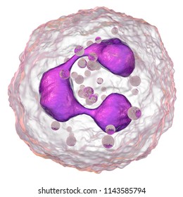
NeutrophilsThe shape of a neutrophil

•         Most numerous WBCs

•         Polymorphonuclear leukocytes (PMNs)

•         Fine granules take up both acidic and basic dyes

•         Give the cytoplasm a lilac color

•         Granules contain hydrolytic enzymes or defensins

Very phagocytic—“bacteria slayers

Eosinophils

•         Red-staining, bilobed nuclei

•         Red to crimson (acidophilic) coarse, lysosome-like granules

•         Digest parasitic worms that are too large to be phagocytized

•         Modulators of the immune response

Basophils

•         Rarest WBCs

•         Large, purplish-black (basophilic) granules contain histamine

•         Histamine: an inflammatory chemical that acts as a vasodilator and attracts other WBCs to inflamed sites

•         Are functionally similar to mast cells

shape of neutrophil, Eosinophil and Basophil

Agranulocytes

•         They includes lymphocytes and monocytes.

•         Lack visible cytoplasmic granules.

•         Have spherical or kidney-shaped nuclei.

Lymphocytes

•         Large, dark-purple, circular nuclei with a thin rim of blue cytoplasm

•         Mostly in lymphoid tissue; few circulate in the blood

•         Crucial to immunity.

•         Two types :T cells act against virus-infected cells and tumor cells B cells give rise to plasma cells, which produce antibodies

Monocytes

•         The largest leukocytes

•         Abundant pale-blue cytoplasm

•         Dark purple-staining, U- or kidney-shaped nuclei

•         Leave circulation, enter tissues, and differentiate into macrophages

•         Actively phagocytic cells; crucial against viruses, intracellular bacterial parasites, and chronic infections

•         Activate lymphocytes to mount an immune response

Composition of Blood and its Functions

2.Red blood cells.( Erythrocytes)

Red blood cells (RBCs), also referred to as red cells, red blood corpuscles, haematids, erythroid cells or erythrocytes are the most common type of blood cell and ...

Function?: ?Oxygen transport

Characteristics of RBCs

  1.  Biconcave discs, no nucleate, essentially no organelles.
  2. Filled with hemoglobin (Hb) for gas transport
  3. Contain the plasma membrane protein spectrin and other proteins
  4. Provide flexibility to change shape as necessary
  5. Are the major factor contributing to blood viscosity

Development of RBC. (Erythropoiesis).

  • The first recognized red cell precursor is called pronormpblast:
  •  Pronormoblast undergoes change in size, colour and contents to reach the mature RBC and passing through the following stages:
Pronormoblast
  1. Early normoblast
  2. Intermediate normoblast
  3. Late normoblast
  4. Reticulocyte
  5. Mature RBC

Red blood cellsBiconcave shape of red blood cell

Structure Human Hemoglobin Molecule Vector Diagram Stock Vector ...

Some reticulocytes enter blood before full maturation
30% of the Hb is formed in the reticulocytes
It takes 2 days to develop into mature RBC
Reticulocyte count in blood is 1%
Reticulocytes count is an indicator of bone marrow activity
An increase in the reticulocyte count (reticulocytosis) indicates increase bone marrow activity e.g. in hemolytic anemia
The mature RBC has no nucleus or other organelles
It is a bag of haemoglobin
A biconcave disc containing haemoglobin
Cells with volume < 80 :microcyte

Cells with volume >95 :macrocyte

Importance of the biconcave shape:
  1. Increases surface area
  2. Increases flexibility
  3. Decreases fragility
  4. Cells with an abnormal shape breaks down easy
  5. Patients with hereditary spherocytosis they have anaemia due to excessive breakdown of the red cell.
  6. Fragility of the cells can be tested by osmotic fragility test

Control of Red Cell Production:
  1. Normal RBC count is 5 million /mm3
  2. Count is higher in males than females. Testosterone enhances EPO production, resulting in higher RBC counts in males.
  3. Red cell function is to transport O2 to tissues
  4. O2 need by the tissues is the main regulator of RBC production
Regulation of RBC production:
Kidney?
Erythropoeitin?  +
Bone marrow?
Red blood cell?
Oxygen?
 
An increase in RBC count is called polycythaemia.
Physiological polycythaemia as occurs at high altitude in response to decrease in O2 pressure
Pathological polycythaemia: as for example a tumor of the kidney producing large amounts of Erythropoeitin
Effects of polycythaemia:- Increases the blood viscosity
Hemoglobin
    1. Found inside the RBC
    2. Main function is O2 transport
    3. Contributes to the buffering capacity of the blood

3. Platelets (Thrombocytes)

Platelets are tiny blood cells that help your body form clots to stop bleeding. If one of your blood vessels gets damaged, it sends out signals to the platelets. The platelets then rush to the site of damage. they form a plug (clot) to fix the damage.

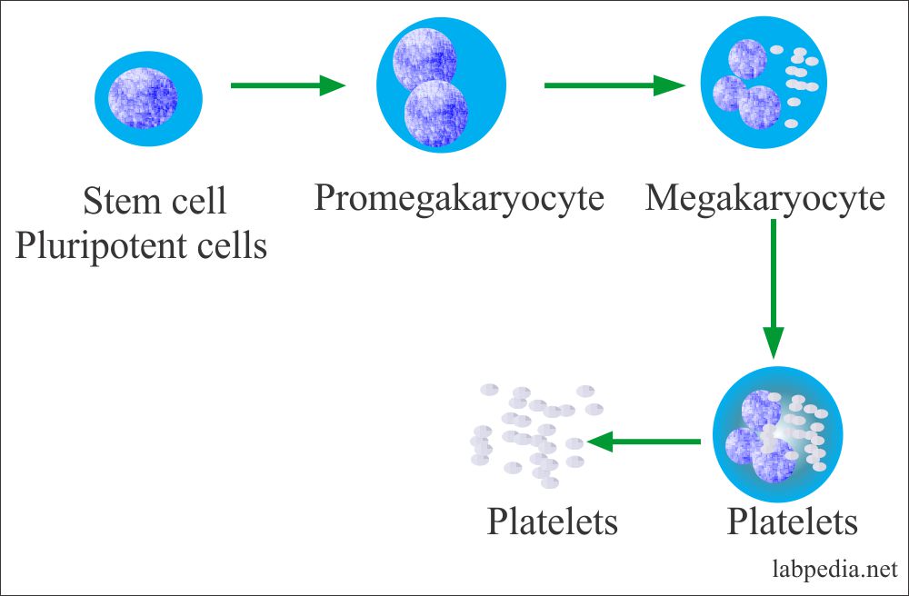
•         They are small fragments of megakaryocytes

•         Formation is regulated by thrombopoietin

•         Blue-staining outer region, purple granules

•         Granules contain serotonin, Ca2+, enzymes, ADP, and platelet-derived growth factor (PDGF

•         Form a temporary platelet plug that helps seal breaks in blood vessels

•         Circulating platelets are kept inactive and mobile by NO and prostacyclin from endothelial cells of blood vessels


Hemostasis( stoppage of bleeding)

•         Fast series of reactions for stoppage of bleeding. Involve:

          1.   -Vascular spasm
          2.   -Platelet plug formation
          3.  - Coagulation (blood clotting

      Vascular spasm

•         Vasoconstriction of damaged blood vessel

•         Triggered by:

    1. -Direct injury
    2. -Chemicals released by endothelial cells and platelets
    3. -Pain reflexes

        Platelet Plug Formation

•          Positive feedback cycle

•         At site of blood vessel injury, platelets stick to exposed collagen fibers with the help of von Willebrand factor, a plasma protein.

•         Swell, become spiked and sticky, and release chemical messengers

•         ADP causes more platelets to stick and release their contents

•         Serotonin and thromboxane A2 enhance vascular spasm and more platelet aggregation

       Coagulation

•         A set of reactions in which blood is transformed from a liquid to a gel

•         Reinforces the platelet plug with fibrin threads.

Three phases of coagulation

    1. •         Prothrombin activator is formed (intrinsic and extrinsic pathways)
    2. •         Prothrombin is converted into thrombin
    3. •         Thrombin catalyzes the joining of fibrinogen to form a fibrin mesh

Platelet - Process of formationPlatelet - Process of formation

                                                                      Platelets

Platelets


Assignment: Read on the three phases of coagulation process


Click here to access Unit Three Content..


Topic 3: Summary

Blood is broadly classified into:

    1. White blood cells (leucocytes)
    2. Red blood cells (erythrocytes)
    3. Platelets (thrombocytes)

1. White blood cells( Leukocytes)

Classification of white blood cells


Granulocytes

Granulocytes: neutrophils, eosinophils, and basophils

 Cytoplasmic granules stain specifically with Wright’s stain

Larger and shorter-lived than RBCs

Lobed nuclei

Phagocytic


Agranulocytes

  1. They includes lymphocytes and monocytes.
  2. Lack visible cytoplasmic granules.
  3. Have spherical or kidney-shaped nuclei.

2.Red blood cells.( Erythrocytes)

Red blood cells (RBCs), also referred to as red cells, red blood corpuscles, haematids, erythroid cells or erythrocytes are the most common type of blood cell and ...

Function?: ?Oxygen transport

Characteristics of RBCs

  1. Biconcave discs, no nucleate, essentially no organelles.
  2. Filled with hemoglobin (Hb) for gas transport
  3. Contain the plasma membrane protein spectrin and other proteins
  4. Provide flexibility to change shape as necessary
  5. Are the major factor contributing to blood viscosity

Composition of blood- summary


Click here to access Unit Three Content..


Topic 4: Blood Typing

Blood typing is a method to tell what type of blood you have. Blood typing is done so you can safely donate your blood or receive a blood transfusion. It is also done to see if you have a substance called Rh factor on the surface of your red blood cells.

Were learned from tragedies (death) caused by mismatch during transfusion in ancient time.

ABO blood types were identified in 1900 by Karl Landstein (1930 Nobel laureate).

Other blood types were identified later.

RBC membranes bear 30 types glycoprotein antigens that are:-

  1. Perceived as foreign if transfused blood is mismatched
  2. Unique to each individual
  3. Promoters of agglutination and are called agglutinogens

NB: Blood type is determined by agglutinogens

Agglutinogens

  • Are specific glycoproteins on red blood cell membranes.
  • All RBCs in an individual carry the same specific type of agglutinogens.
  • Presence or absence of each antigen is used to classify blood cells into different groups
  • On the surface of red blood cells there are antigens
  •  Other blood groups (MNS, Duffy, Kell, and Lewis) are usually weak agglutinogens.

There are 2 types of antigens:

  1. antigen A
  2.  antigen B
  • In the plasma there are antibodies:
  1.  antibody A
  2.   antibody B

 ABO BLOOD GROUP SYSTEM

Type A: RBCs carry agglutinogen A.

Type B: RBCs carry agglutinogen B.

Type AB: RBCs carry both A and B agglutinogens.

Type O: RBCs carry no A nor B agglutinogens.

Accordingly ,the population is divided into four groups:

group                   antigen       antibody

 A                            A                   B          

B                            B                    A

 AB                      A+B                ----

 O                          -----                A+B

Distribution:

        A                                                42%

        B                                                 9%

       AB                                               3%

        O                                                 46%

Inheritance:

Blood group of the individual is determined by 2 genes one from each parent: Mother and father = Baby.

Blood Type Match: D= donor,   R = recipient.

           D

R       

A

B

O

AB

A

Yes

No

Yes?

No

B

No

Yes

Yes

No

O

No

No

Yes

No

AB

Yes

Yes

Yes

Yes


NB: Practice on your own using the table above.

Type AB- Universal recipient

Type O – Universal donor.


Rhesus(Rh)  Blood Group System

The Rh blood group system is one of 36 known human blood group systems. It is the second most important blood group system, after the ABO blood group system. The Rh blood group system consists of 49 defined blood group antigens, among which the five antigens D, C, c, E, and e are the most important.

•         There are 49 different Rh agglutinogens (Rh factors)

•         C, D, and E are most common

•         Rh+ indicates presence of D antigens on the surface of RBCs

•         Rh positive- - RBCs contain Rh agglutinogens.

•         The majority of human beings is Rh positive.

•          Rh negative- - The RBCs contain no Rh agglutinogens..

•         Agglutinins against Rh-positive RBCs are produced after Rh-negative blood sees Rh-       positive RBCs.

•          Anti-Rh antibodies are not spontaneously formed in Rh individuals

•         Anti-Rh antibodies form if an Rh individual receives Rh+ blood

•         A second exposure to Rh+ blood will result in a typical transfusion reaction


Click here to access Unit Four Content..


Topic 1: Structure and Function Terminology

Objectives

By the end of this topic you should be able to;

  1. Demonstrate understanding on the terminology used in muscular system. 
  2. Distinguish the tree types of muscular system 

    Structure and Function Terminology

    Muscle Fibers -muscle cells have multiple nuclei, mitochondria, glycogen granules, lipid droplets, ribosomes, t-tubules, golgi and sarcoplasmic reticulum, (contraction proteins) 

    Sarcomeres -contractile protein units within myofibrils.

    Contractile Proteins -filamentous actin, myosin, tropomyosin, troponin, titin, nebulin

    Striations -visual appearance through electron microscopy of an organized array of light and dark strands within sarcomeres.

    Myofibrils-organized array of sarcomeres connected in series (end to end) along the length of a muscle fiber.

    Sarcomeres - structural units of the myofiber where structural and contractile proteins are organized in a specific sequence, causing a striated appearance under electron microscopy.

    Myosin -the largest of the contractile proteinsS1unit-the globular head region of myosinActin-a globular protein that forms a two stranded filament (F-actin) in vivo

    Tropomyosin -a rod shaped protein attached to actin in a regular repeating sequence.

    Troponin -a 3 component protein that is associated with each actin-tropomyosin complex.

    Sarcolemma -the cell membrane of skeletal muscle.Motor Unit-a single amotor nerve and all the muscle fibers that it innervates

    Excitability –is ability of muscle fibre to receive and propagate an action potential.

    Contractility- is ability of muscle fibre to contract/shorten

    Elasticity- is ability of muscle fibre to rapidly return to a pre-contraction length.  Myofibrils-longitudinal subcellular structures of muscle proteins. 

    The demands of exercise require that skeletal muscles must be able to:

                -contract and generate a wide range of tensions/force,

                -alter tension/force in small increments,

                -do this repeatedly and rapidly for durations that may vary from a few seconds to several hours.

    Functions of Muscles

    Muscles are responsible for movement. The types of movement are locomotionpropulsion of substances through tubes as in circulation, and change in the size of the opening as in the contraction and relaxation of the iris of the eye.

    Muscles help to maintain posture through a continual partial contraction of skeletal muscles in a process known as tonicity.

    Muscles help to produce heat through the chemical changes involved in muscular action.

Click here to access Unit Four Content..


Topic 1: Skeletal Muscle

change management

1. Skeletal Muscle also known as voluntary or muscle fibers that have a striated appearance by the conscious part of the brain. The Muscle fibers re organized into bundles supplied by blood vessels (supplied with the oxygen and glucose needed to fuel contraction)  and innervated by motor neurons and attach to bones by tough, fibrous cords of connective tissue called tendons (connective tissue) or to connective tissues such as ligaments and enable movement. The muscles have a cross-striped appearance, thus striated muscle. There are 600 skeletal muscles that are responsible for the movement of the body. Muscles move through contractility, extensibility, excitability, and elasticity. The process of muscle movement is as follows:

Contractility – allows muscles to change shape to become shorter and thicker.

Extensibility – allows living muscle cells to be stretched and extended; they become longer and thinner. 

Excitability – muscles receive and respond to stimulation.

Elasticity- once the stretching force is removed, a living muscle returns to its original shape. Three distinguishable parts Muscles: the body or main portion, an origin, which is the more fixed attachment, and the insertion, the point of attachment of a muscle to the part that it moves. The means of attachment is a tendon. An Apo neurosis is a wide, thin, sheet like tendon. 

Muscles and nerves work together as a motor unit. They perform in groups and are classified as: Antagonist – a muscle that counteracts the actions of another. Prime Movers or Agonist a muscle that is primary in giving movement. Its contraction produces the movement. Synergist – a muscle that acts with another muscle to produce movement. Skeletal muscles attach to Skeletal muscles are mostly voluntary.

2. Smooth Muscle – also known as involuntary, visceral (airways, stomach, Alimentary canal, and blood vessels), or un striated. Smooth muscle is not controlled by the conscious part of the brain. These muscles are under the control of the autonomic nervous system because it works automatically rather than under conscious control and in most cases, produce relatively slow contractions with greater degree of extensibility. These muscles lack the cross-striped appearance of skeletal muscle. This type of muscles control internal organs of the digestive, respiratory, and urinary tract plus certain muscles of the eye and skin.

Click here to access Unit Four Content..


Topic 1: Cardiac Muscle

3. Cardiac Muscle – the muscle of the heart or myocardium is responsible for circulating blood throughout the body. It has its own pacemaker for rhythmic beating/contracts in response to signals from the cardiac conduction system to make the heartbeat. Cardiac muscle is made from cells called cardiocytes. It is striated in appearance and involuntary because it is controlled by the autonomic nervous system and specialized neuromuscular tissue located within the right atrium.  Pumps blood throughout body– contains more mitochondria than skeletal muscle cells.

Muscular system and the nervous system work together because somatic signals are sent from the cerebral cortex to nerves associated with specific skeletal muscles. Most signals travel through spinal nerves that connect with nerves that innervate skeletal muscles throughout the body.

Muscle contraction begins when the nervous system generates a signal (an impulse) called an action potential, travels through a type of nerve cell called a motor neuron. The neuromuscular junction is the name of the place where the motor neuron reaches a muscle cell. Skeletal muscle tissue is composed of cells called muscle fibers.

 Brain neuron sending an electronic signal to a motor neuron.

 When the nervous system signal reaches the neuromuscular junction a chemical message is released by the motor neuron. The chemical message, a neurotransmitter called acetylcholine, binds to receptors on the outside of the muscle fiber. That starts a chemical reaction within the muscle, to make it contract. A multistep molecular process within the muscle fiber begins when acetylcholine binds to receptors on the muscle fiber membrane. 

The proteins inside muscle fibers are organized into long chains that can interact with each other, reorganizing to shorten and relax. When acetylcholine reaches receptors on the membranes of muscle fibers, membrane channels open and the process that contracts a relaxed muscle fibers begins.

When the stimulation of the motor neuron providing the impulse to the muscle fibers stops, the chemical reaction that causes the rearrangement of the muscle fibers proteins is stopped. This reverses the chemical processes in the muscle fibers and the muscle relaxes.

Sliding Filament Theory of muscular contraction explains how cross-bridges are formed by actin and myosin filaments within the sarcomeres of muscle fibres bind to create cross-bridges and slide past one another, creating a contraction. 

For a contraction to occur there must first be a stimulation of the muscle in the form of an impulse (action potential) from a motor neuron (nerve that connects to muscle). Then an impulse stimulates the 'heads' on the myosin filament to attach to the actin filament and pull actin towards the centre of the sarcomere resulting to the shortening of all sarcomeres. This causes Troponin attached to the protein tropomyosin within the actin filaments. 

When the muscle is relaxed tropomyosin blocks the attachment sites for the myosin cross bridges (heads), thus preventing contraction.

When the muscle is stimulated to contract by the nerve impulse, calcium channels open in the sarcoplasmic reticulum (which is effectively a storage house for calcium within the muscle) and release calcium into the sarcoplasm (fluid within the muscle cell).  Some of this calcium attaches to troponin which causes a change in the muscle cell that moves tropomyosin out of the way so the cross bridges can attach and produce muscle contraction.


Click here to access Unit Four Content..


Topic 1: Summary

Summary the sliding filament theory of muscle contraction can be broken down into four distinct stages, these are;

1. Muscle activation: the motor nerve stimulates an action potential (impulse) to pass down a neuron to the neuromuscular junction. This stimulates the sarcoplasmic reticulum triggering release of calcium into the muscle cell.
 
2. Muscle contraction:  Calcium floods into the muscle cell binding with troponin allowing actin and myosin to bind.  The actin and myosin cross bridges bind and contract using ATP as energy (ATP is an energy compound that all cells use to fuel their activity. 
 
3. Recharging:  ATP is re-synthesised (re-manufactured) allowing actin and myosin to maintain their strong binding state
 
4. Relaxation:  Relaxation occurs when stimulation of the nerve stops.  Calcium is then pumped back into the sarcoplasmic reticulum breaking the link between actin and myosin. Actin and myosin return to their unbound state causing the muscle to relax.  Alternatively relaxation (failure) will also occur when ATP is no longer available.

In order for a skeletal muscle contraction to occur;

1.There must be a neural stimulus
2. There must be calcium in the muscle cells
3. ATP must be available for energy

Click here to access Unit Four Content..


Topic 2: The Heart

Anatomy of the heart- Overview

Size: Size of a closed fist

Shape:  Apex: Blunt rounded point of cone

  Base: Flat part at opposite of end of cone

Location: Located in thoracic cavity in mediastinum

Surrounded and cushioned by the lungs

Heart size = approx. 300gms

Heart chambers:

Right atrium and ventricle

Left atrium and ventricle

Atrium ? receives blood from veins

Ventricles ? eject blood into arteries

Blood supply: supplied by right and left coronary arteries

Nerve supply: Control is by sympathetic system and the vagus nerve


Functions of the Heart

  1. Generating blood pressure 
  2.   Routing blood -Heart separates pulmonary and systemic circulations
  3. Ensuring one-way blood flow-Heart valves ensure one-way flow
  4. Regulating blood supply- Changes in contraction rate and force match blood delivery to changing metabolic needs.

Physiology of cardiac muscle

The 3 major types of heart muscle:

?      The atrial muscle

?      The ventricular muscle

?      The specialized excitatory and conductive muscle fibres

Atrial and ventricular muscles contract just the same way as skeletal muscle but the duration is much longer. This gives rise to mechanical activity of the heart i.e contraction and relaxation of the two muscles.

The specialized excitatory and conductive muscle fibre.This muscle exhibits automatic rhythmical electrical discharge inform of action potential. It controls the rhythmical beating of the heart i.e electrical activity of the heart.


Physiology of cardiac muscle cont’d

Conducting system (specialized excitatory and conductive muscle)- controls and coordinates heartbeat

Contractile cells(atrial and ventricular muscles)- produce contractions

The Cardiac Cycle begins with action potential at SA node and:

  1.  transmitted through conducting system
  2. produces action potentials in cardiac muscle cells (contractile cells)

Electrical events in the cardiac cycle can be recorded on an electrocardiogram (ECG)


Superficial Anatomy of the Heart

4 cardiac chambers

Atria - Thin-walled

Expandable outer auricle

Coronary sulcus- divides atria and ventricles

Anterior and interventricular sulci -separate left and right ventricles

                                                               -contain blood vessels of cardiac muscle

Heart valves

CLICK NEXT

Hero media Hero media
order by votes Up vote 18 Down vote Instead of using a plugin with unnecessary Kbytes, all you need is a simple function like this (see explanation in comments):